General Instructions:
- Copy the given assignment of practical in fair practical copy of biology.
- Make the use of black pen for heading and sub-headings and blue pen for writing other content.
- Work should be presentable.
- Make well labelled and proper diagram as provided in this assignment.
Spot No.7
Aim: To study and identify Ascaris and symptoms of disease caused by it
Materials Required: Museum specimens of Ascaris, Compound microscope
Procedure:
- Observe the museum specimen of Ascaris carefully.
- Draw a well labelled diagram of Ascaris observed.
Observations:
Classification
Phylum: Nemathelminthes
Class: Nematoda
Order: Ascaroidea
Genus: Ascaris
Species: lumbricoides
Comments:
- Ascaris is endoparasite of man found in small intestine.
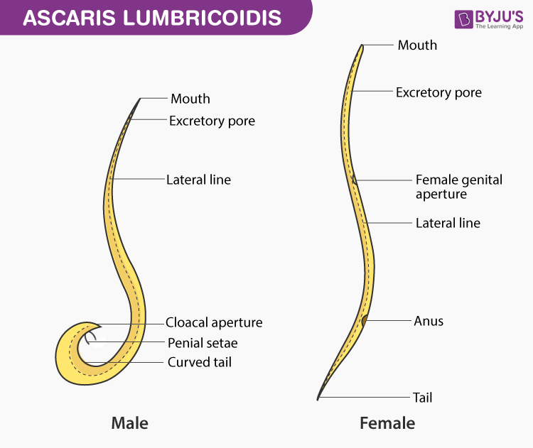
- It is elongated, cylindrical, wormiform, yellowish or pinkish in colour and tapering at both the ends.
- Size.
- Male: 15-30cmX3-5mm
- Female: 20-40 cmX 6-8 cm
- Mouth is triradiated at anterior end and surrounded by 3 lips (1 mid dorsal + 2 ventrolateral).
- It possess 4 longitudinal epidermal lines in the entire length of the body (1 dorsal, 1 ventral and 2 lateral lines).
- Amphid (chemoreceptor) gustatory helps in feeding.
- Possess well marked sexual diamorphism,
- Tail is curved ventrally curved in males while straight in females.
- Male is smaller than females.
- Genital aperture in females is at mid ventral from anterior end.
- Penial spicule, cloaca, pre-anal and post-anal papillae are present in male but absent in female.
- Life span of Ascaris is 9-12 montghs
- It causes Ascariasis disease.
Mode of infection
- Food and water contaminated with embryonated eggs
- Second stage rhabditiform larva is infective stage of parasite.
Symptoms of Ascariasis
- Loss of appetite
- Insomnia
- Colic pain
- Abdominal discomfort
- Irregular bowel movement
- Occasional vomiting
- Anaemia
Prrecautions:
- Handle the museum Specimen carefully.